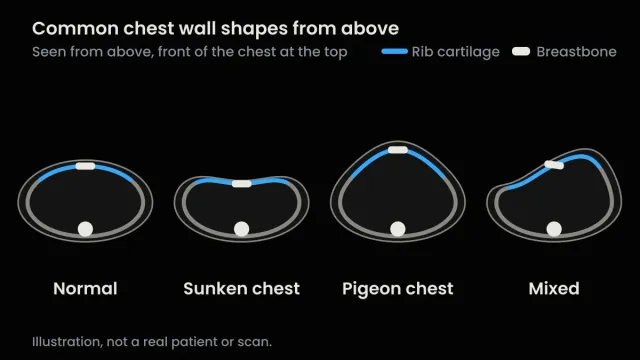
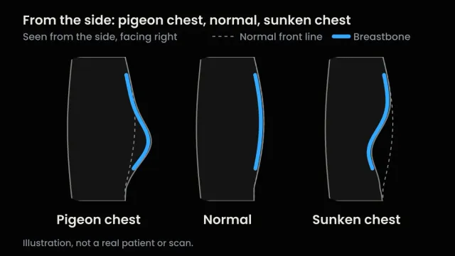
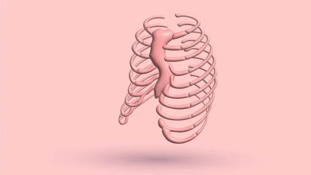
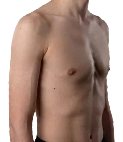
Contents · 10 sections
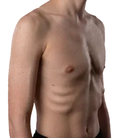
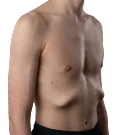
Your scan report says something like “Haller index 3.4”, and you want to know whether that’s bad. The short answer is that the Haller index is a ratio: the inside width of your chest divided by the shortest gap between your breastbone and your spine. About 2.5 is average for a chest without pectus excavatum. Above 3.25 is a common line for a surgical opinion and for insurance approval. It isn’t an emergency, and it doesn’t decide surgery on its own.
It’s also less fixed than it looks. Breathing and scan type can move it, and it describes shape, not how your heart and lungs are working. This guide covers how it’s calculated, how to read yours, why scans disagree and what to ask your doctor.
I was diagnosed with pectus excavatum at 15 and corrected it without surgery over about three years. The change was visible and measurable, but not complete. I’m not a doctor, so every medical point here comes with the study, guideline or policy behind it. If you’re new to the condition itself, start with my guide to what pectus excavatum is.
What the Haller index measures
The index comes from one cross-section of a chest scan, called an axial slice: the view you’d get if the chest were cut straight across, like a loaf of bread. On the slice where the dip is deepest, two distances are measured:
- A: the widest inside width of the chest, from the inner edge of the ribs on one side to the other.
- B: the shortest distance from the back of the breastbone to the front of the spine.
A is divided by B. The units cancel, so the index is a plain number. A deeper dip shrinks B, so the index goes up.
The ratio comes from a 1987 preliminary report by Haller, Kramer and Lietman, who used CT to help select children and teenagers for surgery. You’ll also see it called the pectus index. In their series, every patient who needed an operation was above 3.25 and every normal comparison chest was below it. That small report is where the 3.25 line comes from.
A worked example
These numbers are made up for illustration, not taken from real patients. They just show the arithmetic:
| Inside width (A) | Breastbone to spine (B) | Haller index |
|---|---|---|
| 26 cm | 10.4 cm | 2.5 |
| 26 cm | 8 cm | 3.25 |
| 26 cm | 6.5 cm | 4.0 |
| 30 cm | 8 cm | 3.75 |
Look at the last row. The gap behind the breastbone is the same 8 cm as in the second row, but the chest is wider, so the index is higher. Same dip, different number, just because one chest is broader.
Work out the ratio from your scan report
Enter the two measurements from a CT or MRI report, in the same unit. A ruler on your chest at home can't give you these.
Your ratio appears here: A divided by B.
No single number decides surgery. What to do with your number.
What your number means
About 2.5 is average, not a target
In a Children’s Mercy Kansas City study, 220 people without pectus excavatum averaged 2.35 on CT, against 4.06 in 252 who had surgery. An 11-center surgical study and Cigna’s coverage policy both give 2.5 as normal. So “about 2.5” is a fair average, but healthy chests don’t all hit it.
The index also changes with age and with the level of the chest where it’s measured. In normal CT scans of 8- to 18-year-olds in the UK, it was highest near the top of the chest, and that upper part changed with age. The authors concluded that one normal range can’t fit every age and every level. For parents, there’s more in my guide to sunken chest in babies and children.
3.25 is a line for a surgical opinion, not an emergency
A Haller index around 2.5 is average and 3.25 or more is usually called severe, but no single number decides surgery. The 2024 Pectus Care Guidelines, a consensus from several surgical and Marfan societies, say 3.25 is consistently used to mark the severe category. Crossing it doesn’t mean something urgent is happening, and it isn’t enough on its own:
- In the guidelines, the clearest case for surgery is an index above 3.25 together with objective evidence that the heart is being compressed. They also describe most cases as mild and well tolerated.
- At the Norfolk, Virginia, center where the Nuss procedure was developed, 51% of 2,378 people evaluated went on to have surgery. They needed symptoms, a clinically severe chest and at least two objective findings, and an index over 3.25 was only one of those. The others were heart or lung compression, a heart valve problem, an abnormal heart rhythm and restricted lung function.
And 3.25 isn’t sacred. In a 2018 viewpoint, Eisinger and Pilegaard argued the cut-off has never been validated and that insurers should weigh more than the index. It’s an opinion piece, not proof, but a fair warning against treating 3.25 as a law of nature.
Mild and moderate are labels, not a standard
Mild and moderate have no agreed cut-offs. The 2024 guidelines define only severe, at 3.25 or more. The 11-center study gives a normal value (2.5) and a severe line (above 3.2), nothing in between. So treat any mild or moderate ranges you see quoted as rough labels, and if your report says “moderate”, ask which scale it’s using. The two numbers worth anchoring to are about 2.5 and 3.25.
A higher number means a harder repair, not a more urgent one
If you do have surgery, the index matters in another way. Among 2,013 people who had the Nuss procedure in Denmark, 16.4% had a complication. Each extra point of Haller index raised the odds by about 10%, and at 5 or more, complications were 2.2 times as common. A higher number tells you how hard a repair may be, not how soon you need one.
Why two scans can give different numbers
Several things change the index without your chest changing at all.
Breathing in or out
The index reads higher at the end of a breath out than at the end of a breath in, by more than most people expect.
- In an MRI study of 47 patients at Cincinnati Children’s (median age 14), the median index was 3.96 on a breath in and 5.09 on a breath out. Of the 11 patients below 3.25 on the breath in, 8 were above it on the breath out.
- In a CT study of 36 patients in Buenos Aires, the share who met surgical criteria rose from 71% on the breath in to 91% on the breath out.
- The 2024 guidelines say the index can be up to 30% higher at the end of a breath out.
This matters most near the line. In one real-time MRI study of 56 patients with a median index of 5.7, only 4 crossed 3.25 between a full breath in and out, as a 2024 review of pectus imaging points out, because most were far above it. The Buenos Aires team and the real-time MRI team both recommend scanning at the end of a breath out, but centers differ. Ask which phase your scan used, especially if you’re close to 3.25.
CT, X-ray or MRI
CT is the reference. Besides the index, it shows whether the heart is compressed or pushed aside, how lopsided the chest is and whether the breastbone is twisted, according to the 2024 imaging review. A full chest CT is about 1 to 2 mSv (millisieverts, the unit for radiation dose), and low-dose pectus protocols can bring that to about 0.35 mSv or less.
A chest X-ray from the front and side is about 0.02 mSv. It’s least reliable right where it matters. In a study of 77 patients in Iowa, the X-ray index rarely missed a chest that CT called severe, but it also flagged some that CT put below the line. The authors suggest confirming with CT when the X-ray index falls between 3.2 and 3.75. The imaging review adds that X-ray isn’t suitable for markedly lopsided chests.
MRI uses no radiation and can also show how the heart is working. In a Beijing study of children, its index wasn’t significantly different from CT’s.
The level of the chest matters too. As the UK study above showed, the index changes from one level to another, and the level used isn’t standardized, which is one of the points Eisinger and Pilegaard made. All three scans are usually done lying on your back. Standing can change the number: in a Japanese study using upright and lying CT, the index of 21 people with pectus was higher standing than lying (median 10.1 against 6.4), mostly because the lower spine shifted forward, while 35 volunteers without pectus barely changed (2.9 against 2.8).
The practical rule: compare like with like, meaning the same scan type, breathing phase, level and ideally center. Otherwise a change in your number may just be a change in method.
What the Haller index can’t tell you
If you’re worried about your heart or lungs, this is the part that matters. The index describes shape on one slice, and what you probably care about lines up with it only loosely.
- How you feel. In the PCAPES study of 432 adults referred for surgery, symptom scores barely tracked the index: a correlation of 0.16, where 0 means no link and 1 a perfect one. All were surgical referrals, so it says less about milder chests.
- Whether your heart is squeezed. Compression shows up on the images, and you can’t work it out from the number. In 124 people aged 10 to 19 evaluated before surgery in Memphis, 24% had PVCs (premature ventricular contractions, early extra beats from the heart’s lower chambers) during an exercise test. They were linked to the right side of the heart being compressed on CT, not to the Haller index.
- Your fitness, exactly. In a German study of 99 otherwise healthy patients, mostly young, heart rate at the same point of hard effort (the anaerobic threshold) was higher above 3.25, but peak oxygen uptake, the standard measure of aerobic fitness, was lower only above 4.0.
- The full shape of your chest. Width changes the index, as the worked example showed. It also ignores how lopsided the chest is, whether the breastbone is twisted and how far the dip runs up and down. In the Children’s Mercy study, Haller values showed a 48% overlap between the pectus and normal groups, while correction index values didn’t overlap at all.
- Sex differences. In 345 patients evaluated before surgery in Cincinnati, 19% of them female, the girls and women had higher indices but better heart function and exercise results than the males.
So doctors don’t judge a chest by the ratio alone. They look at the images for compression and use functional tests: lung function tests (how much air you can move), an echocardiogram (an ultrasound of the heart) and an exercise test. There’s more in my guides to heart problems with pectus excavatum and pectus excavatum later in life.
Other numbers you may see on your report
| Index | What it measures | What it’s used for |
|---|---|---|
| Correction index | How much of the chest’s front-to-back depth the dip takes away, as a percentage | 28% or more corresponds to a Haller index of 3.25. In 75 patients, it held up in unusually shaped chests where the Haller index didn’t |
| Asymmetry index and sternal tilt | How lopsided the chest is, and how far the breastbone is tilted or twisted | What the Haller index ignores. Neither changed with breathing in the Cincinnati MRI study |
| Cardiac compression index | The width of the heart divided by the breastbone-to-spine gap | How squeezed the heart is. It also rises on a breath out |
| Titanic index | How much of the breastbone’s length sits behind the line of the front ribs | Surgical planning. In 78 patients, above 66.5% predicted needing more than two bars in a Nuss procedure |
Many more exist, according to the 2024 imaging review, and papers don’t always define them the same way. A “depression index”, for example, means different things in different studies. If your report names one you can’t place, ask what it means for you.
How insurers use the Haller index
In the policies I could check, 3.25 is one condition for paying for surgery, never the only one. Policies change, so here they are with dates.
| Who | Haller rule | What else is needed | Date |
|---|---|---|---|
| Aetna, policy 0272 (US) | Above 3.25, on CT | At least one of: heart compression or displacement with reduced output on an echocardiogram, total lung capacity at or below 80% of predicted, or objective exercise intolerance. Otherwise it’s classed as cosmetic | Last reviewed 17 Apr 2026 |
| Cigna, policy 0309 (US) | Above 3.25, on imaging such as CT, X-ray or MRI | Lung function tests showing restrictive or obstructive lung disease, or heart imaging showing compression. Coverage varies by plan | Effective 15 Apr 2026 |
| NHS England interim policy | Above 3.25 | A national specialist team confirming potentially life-threatening complications, plus one of: an abnormal heart rhythm, fainting or near-fainting, heart compression on MRI with symptoms, trouble swallowing, or a chest that blocks other surgery. About 5 to 10 patients a year expected | File dated Apr 2023 |
Aetna names CT while Cigna also lists X-ray and MRI, so the scan you had can matter. Both want a functional finding on top of the number.
Children’s Mercy Kansas City’s coverage sheet for families, reviewed in May 2026, says approval typically requires an index above 3.25, and some insurers also want an exercise test, lung tests or an echo showing a problem. If those come back normal, the insurer may deny coverage even above 3.25, and you may need to appeal.
Get your plan’s written policy, and ask the surgeon’s office which results they’ll send. Costs and appeals are in my guide to pectus surgery cost and appeals.
Can you work out your Haller index at home?
No. Both distances are measured inside the chest, on a scan.
Home calculators that divide your chest’s width by its depth, measured on the skin, give a number that looks like a Haller index but runs on a lower scale. In 197 children and teens aged 4 to 18, the skin-to-skin caliper ratio that best picked out those who went on to surgery was 1.83, against 3.25 on CT. Compare a home ratio with 3.25 and your chest will look milder than a scan would say. 3D surface scanning looks promising, but the imaging review says it isn’t standardized yet and is less accurate in people with breasts or more body fat.
What you can do at home is measure the depth of the dip the same way every month and take photos. A ruler reading is your tracking number. It is not a Haller index. My guide to measuring pectus excavatum at home shows how.
Does treatment change the number?
Surgery does. In the 11-center study above, the average CT index fell from 4.4 before repair to 3.0 after.
The vacuum bell is murkier, and the results differ by age:
- In 47 children (median age 7) scanned before and after a year, the average index fell from 3.6 to 3.2. Fat in front of the breastbone thickened too, and the surface dip improved more than the bone did (4.0 mm against 1.4 mm).
- In 19 male patients (median age 14.8) after at least a year, the index didn’t change significantly (5.4 to 5.3). Fat over the dip grew by 6.5 mm, and the authors put the visible improvement mainly down to that.
So part of the flatter look after the bell can come from soft tissue rather than the breastbone moving, and in the teenage study it was most of it. The full evidence is in my guide to vacuum bell therapy.
I found no study showing that exercise, breathing or posture work changes the Haller index. That isn’t proof it can’t. Training works on the muscle and posture around the dip, which my guide to pectus excavatum exercises covers.
What to do with your number
Pick the line that sounds like you.
- Any red flag from the list below. See a doctor before any device, brace or exercise program.
- Above 3.25 with symptoms. Ask for an assessment with a surgeon who treats chest wall conditions. The number itself isn’t an emergency, and you can still decide what to do afterwards.
- Close to 3.25, either side. Ask which breathing phase and scan type were used, and whether a correction index or heart imaging changes the picture.
- A mild chest and no symptoms. A mild, symptom-free chest doesn’t usually need a routine CT scan just to find out the number. To see which route fits you, take my free 1-minute quiz. It has a “not sure” option if you haven’t been measured or assessed.
- You want to try without surgery. Every non-surgical route is on the pectus excavatum treatment without surgery page. Give it honest time. Structural change is measured in months and years.
See a doctor first if any of these apply:
- chest pain
- fainting or nearly fainting
- breathlessness at rest or with light effort
- palpitations or an irregular heartbeat
- a diagnosed heart or lung condition
- Marfan syndrome or Ehlers-Danlos syndrome, or a doctor has raised either
- previous chest surgery
- a Haller index above 3.25 together with symptoms
- symptoms that nobody has checked yet
Questions to ask the doctor who ordered your scan
- Was the scan taken at the end of a breath in or a breath out?
- Was it CT, X-ray or MRI, and at what level of the chest was the index measured?
- What’s my correction index?
- Do the images show my heart being compressed or pushed aside? Is my breastbone twisted?
- Do I need lung function tests, an echocardiogram or an exercise test?
- If surgery comes up, what does my insurance plan require, and which results will you send?
Sources
- Haller JA Jr, Kramer SS, Lietman SA. J Pediatr Surg. 1987;22(10):904-6. Read it
- St Peter SD, et al. J Pediatr Surg. 2011;46(12):2270-3. Read it
- Kelly RE Jr, et al. J Am Coll Surg. 2013;217(6):1080-9. Read it
- Archer JE, et al. J Anat. 2016;229(4):577-81. Read it
- Dunning J, et al. Pectus Care Guidelines. Eur J Cardiothorac Surg. 2024;66(1):ezae166. Read it
- Kelly RE, et al. Ann Surg. 2010;252(6):1072-81. Read it
- Eisinger RS, Pilegaard HK. Biomed J Sci Tech Res. 2018;2(1). Read it
- Media AS, et al. J Thorac Cardiovasc Surg. 2024;168(3):699-711. Read it
- Birkemeier KL, et al. AJR Am J Roentgenol. 2011;197(5):W934-9. Read it
- Albertal M, et al. J Pediatr Surg. 2013;48(10):2011-6. Read it
- Coorens NA, et al. J Thorac Dis. 2024;16(1):696-707. Read it
- Poston PM, et al. Innovations (Phila). 2014;9(2):117-21. Read it
- Sun J, et al. Pediatr Investig. 2019;3(2):102-9. Read it
- Jaroszewski DE, et al. J Thorac Dis. 2024;16(12):8565-81. Read it
- Guerrier K, et al. Med Sci (Basel). 2026;14(3):379. Read it
- Abu-Tair T, et al. Ann Thorac Surg. 2018;105(2):455-60. Read it
- Casar Berazaluce AM, et al. Pediatr Surg Int. 2020;36(11):1281-6. Read it
- Poston PM, et al. Ann Thorac Surg. 2014;97(4):1176-9. Read it
- Bellía-Munzón G, et al. J Pediatr Surg. 2023;58(4):605-7. Read it
- Clinical Policy Bulletin 0272 (pectus excavatum and pectus carinatum). Aetna. Last reviewed 2026 Apr 17 [cited 2026 Oct 20]. Read it
- Surgical treatment of chest wall deformities. Medical Coverage Policy 0309. Cigna. Effective 2026 Apr 15 [cited 2026 Oct 20]. Read it
- Interim clinical commissioning urgent policy statement: pectus surgery (PR00394), v0.2. NHS England. 2023 Apr [cited 2026 Oct 20]. Read it
- Pectus excavatum surgery: understanding your coverage. Children’s Mercy Kansas City. Reviewed 2026 May 26 [cited 2026 Oct 20]. Read it
- Chen T, et al. Front Pediatr. 2022;10:1015026. Read it
- Zhang Q, et al. J Pediatr Surg. 2026;61(7):163131. Read it
- Feng X, et al. JPRAS Open. 2026;48:404-14. Read it
- Suzuki T, et al. Eur J Cardiothorac Surg. 2024;66(5):ezae408. Read it
Reading about the condition is one thing. Knowing which one you have, how severe it is and what to do first takes about a minute.
- Your condition named, not guessed
- A severity read you can act on
- The exact next step for your case

This article is for educational purposes only and does not constitute medical advice. Always consult a qualified healthcare professional before starting any treatment plan. View full disclaimer →