Contents · 12 sections
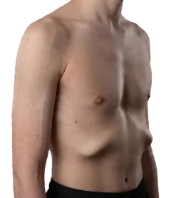
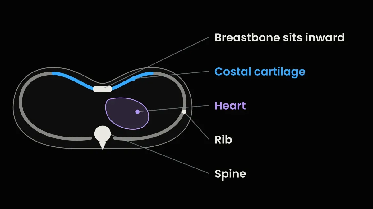
You’ve noticed the dip, in your own chest or your child’s, and the first question is why. The honest answer is that nobody knows the single cause. The problem seems to sit in the costal cartilage, the flexible strips that join the ribs to the breastbone. It often runs in families. A minority of cases come with a genetic syndrome or after specific events such as heart surgery. And nothing you did caused it.
Plenty of pages state some causes as settled when they aren’t. So below, every suggested cause gets a grade for its evidence, and every number comes with the people it was measured in. New to the condition? Start with what pectus excavatum is.
I was diagnosed with pectus excavatum at 15 and corrected it without surgery over about three years. The change was visible and measurable, but not complete. I’m not a doctor, so everything clinical here comes from published studies and hospital sources.
Every cause at a glance
The grades are my own reading of the research, not an official scale.
- Well documented: several studies agree.
- Mixed: studies point both ways.
- Early or small: single families, small series or animal work.
- Abandoned or unrelated: tested and dropped, or not found in people with pectus.
- No study found: I couldn’t find research that tested it.
| Suggested cause | Grade | Verdict |
|---|---|---|
| Family history | Well documented | Roughly 4 to 5 in 10 patients have an affected relative |
| A specific gene | Early or small | About 14 candidates, mostly from single families and not repeated |
| Cartilage growing too long | Mixed | The most popular theory; scan studies disagree |
| Weak cartilage, rib overgrowth, breastbone growth | Early or small | Plausible, not shown |
| Diaphragm pulling the breastbone in | Abandoned or unrelated | Tested and dropped |
| Rickets or low vitamin D | Abandoned or unrelated | Older studies found no signs of it in people with pectus |
| Growth spurt | Well documented | Explains when it worsens, not why |
| Marfan, Noonan, Loeys-Dietz | Well documented | Common in these syndromes; most people with pectus don’t have one |
| Turner syndrome | Early or small | A broad chest is typical; true pectus in a few |
| Ehlers-Danlos syndrome | Early or small | Reported more often; most people with pectus don’t have EDS |
| Klinefelter syndrome | No study found | One case report; no measured link |
| Open-heart surgery | Early or small | Can appear or worsen afterwards |
| Diaphragmatic hernia at birth | Mixed | More pectus in some series, not in others |
| Blocked airway or SMA type 1 in infancy | Early or small | Small studies and case reports |
| Premature birth | No study found | Only a different rib pattern has been studied |
| Posture | No study found | Common alongside pectus; not shown as a cause |
| Weight, exercise, sleep, smoking | No study found | Not tested, as far as I could find |
It runs in families, but the numbers are easy to misread
Family history is the best-supported finding there is. About 1 to 8 people per 1,000 have pectus excavatum, and it’s more common in boys, according to Cleveland Clinic. Cleveland adds that 40% to 53% of people with it have a family member with the same condition, without saying who was counted.
Studies that name their patients land in the same range. A 2022 Hamburg study surveyed patients with isolated pectus excavatum, meaning no known syndrome: 54% of the 78 with family data had an affected relative. In 206 patients operated on in Istanbul and followed up by phone, 47% reported a family history. That study was co-authored by Prof. Yüksel, the site’s medical reviewer.
How it’s passed down is less clear. Dominant means one copy of a gene change from one parent is enough; recessive means it takes a copy from each parent; X-linked means the gene sits on the X chromosome and tends to show up more in boys. A 2006 study of 34 US families found 14 that looked dominant, 4 recessive, 6 X-linked and 10 more complex, and concluded most are probably multifactorial: several genes plus other influences. A 2012 analysis of 48 family trees from the same Norfolk, Virginia, centre pointed mainly to recessive inheritance instead. The studies disagree.
They do agree on the boy bias. In the Istanbul group 85% were male, and so were five in six of 1,046 Danish surgical patients.
Will my children get it?
This is the part that’s easy to get backwards. “About half of patients have an affected relative” does not mean “a child of a parent with pectus has a 40-50% chance”. The first number looks back at relatives of people who already have it. It doesn’t count how many children of affected parents develop it. An earlier version of this page made that mistake.
I couldn’t find a study that gives a child’s risk. A 2014 clinical genetics review from Amsterdam says the risk for isolated pectus with no family history is unknown but thought to be low.
Is pectus excavatum genetic? What gene studies found
Genetics is part of the story, but no single gene has been found. The most recent summary I found, a 2026 UK systematic review, covers studies up to November 2025.
It found about 14 candidate genes or gene regions. Most were rare variants from a single family or case report, and most haven’t been repeated. Every study it included was done in European or East Asian people. And nobody has yet run a GWAS of isolated pectus excavatum: a scan comparing the genes of thousands of people with and without the condition.
The cartilage theory, and why it’s contested
The most common explanation is that the costal cartilage grows too long and buckles the breastbone inward. The Nuss procedure was designed around it, and a 2024 Dutch review still calls overgrowth or disturbed cartilage growth the leading hypothesis, while noting it’s controversial.
When researchers measured the cartilage on scans, results split:
- Against: Japanese CT studies found the cartilage on the deeper side shorter or no different, and the cartilage-to-rib ratio no larger than in people without pectus. A Florida study of 16 patients and 16 matched controls found cartilage was shorter relative to bone. And rats given a hormone that made their cartilage overgrow didn’t develop pectus, a 2022 systematic review notes.
- For a length problem: a Korean study of 19 adults found longer ribs, not longer cartilage. A 2020 Japanese study found the combined rib-and-cartilage length was longer than normal.
So the cartilage is probably involved, but “it grows too much” is a disputed theory, not a fact. The 2022 review lists other early ideas: weaker cartilage, ribs that grow too long, and disturbed growth of the breastbone itself. All plausible, none shown.
Theories that have been dropped
The oldest idea was that the diaphragm pulls the lower breastbone inward. It was abandoned: the deepest point of the dip usually sits above where the diaphragm attaches, and an operation designed to cut that pull didn’t correct the chest.
Rickets also gets listed, including by MedlinePlus. But older studies found no signs of rickets in people with pectus excavatum, and the 2022 review lists vitamin D as unrelated.
Why it often appears in the growth spurt
The growth spurt is when pectus excavatum tends to show up or deepen. Surgeons at a large Norfolk, Virginia, centre write that only 22% of cases are noticed in the first decade of life. At puberty a mild dip can become severe within 6 to 12 months, in their experience.
Researchers think fast skeletal growth may expose a mismatch between the ribs, cartilage and breastbone, but that hasn’t been demonstrated. The full timeline by age, including the adults whose dip keeps deepening, is in does pectus excavatum get worse with age.
Syndromes linked to pectus excavatum
Some genetic syndromes include a sunken chest, but most people with pectus don’t have a syndrome. Both are true because the numbers run in two directions: “how many people with Marfan have a pectus deformity” and “how many people with pectus have Marfan” have very different answers.
| Condition | Pectus in people with the condition | The condition in people with pectus |
|---|---|---|
| Marfan syndrome | A pectus deformity, sunken or pigeon, in up to about 70% (UK review) | 0.4% of 1,046 surgical patients (Denmark); 2.8% of 1,215 (US); 5.3% of 241 referred children (New York) |
| Loeys-Dietz syndrome | 35 of 65 (54%), one US hospital | No figure found |
| Noonan syndrome | 54% of 107 gene-confirmed patients (South India) | 2 of 18 children under 11 at one Dutch clinic |
| Turner syndrome | Mild pectus in 3 of 15 (US, 1993) | No figure found |
| Ehlers-Danlos syndrome | 3.7% self-reported (hypermobile EDS survey); 6 of 20 in one small series | 2 of 1,046 surgical patients (Denmark) |
| Klinefelter syndrome | No study found | No study found |
Marfan syndrome
Marfan is a connective tissue condition caused by changes in the FBN1 gene. It affects about 1 in 5,000 people, and at least a quarter of cases come from a new gene change rather than a parent, according to MedlinePlus Genetics. A UK review puts pectus deformity at up to about 70% of people with Marfan. That figure mixes sunken and pigeon chest, so it isn’t a pectus excavatum rate. In the revised Ghent criteria doctors use to score Marfan, pigeon chest even earns 2 points and sunken chest 1.
The other direction depends on who’s screened. Marfan was found in 0.4% of the Danish surgical patients, 2.8% of US surgical patients, and 5.3% of 241 New York children who were routinely sent for genetic and heart checks (that group included pigeon chests). The 2024 Pectus Care Guidelines estimate about 5% of people with apparently isolated pectus are later diagnosed with Marfan. Marfan can affect the heart and the aorta, the body’s main artery, so a diagnosis is worth having.
Noonan, Loeys-Dietz and Turner
In 107 people with gene-confirmed Noonan syndrome across four hospitals in South India, 54% had pectus excavatum. Loeys-Dietz showed the same rate in a Johns Hopkins series: 35 of 65 patients, and 16 of those 65 had first been diagnosed with Marfan. In Turner syndrome a broad “shield” chest is typical, and a 1993 study of 15 girls found mild pectus excavatum in 3.
Ehlers-Danlos syndrome
Pectus excavatum is reported more often in some types of Ehlers-Danlos syndrome (EDS), mostly in small studies and one survey. Most people with pectus excavatum don’t have EDS. In a global online survey, 125 of 3,360 people with hypermobile EDS (3.7%) said they had pectus excavatum, against 0.1% in an unmatched comparison group. A 2021 review cites a small series in which 6 of 20 people with classical or vascular EDS had mild pectus. That’s an association, not a cause, and an earlier version of this page overstated it.
Klinefelter syndrome
I couldn’t find a study measuring how often pectus excavatum occurs in Klinefelter syndrome. When I searched PubMed in October 2026, it had just two records mentioning both: a 2004 case report and a 2020 Dutch series on tall children in which the child with pectus had Marfan, not Klinefelter. Klinefelter affects about 1 in 450 to 600 male births, according to a 2026 review, and usually makes boys tall. My reading: shared tall stature is the likely reason they get linked. If you have both, tell your doctor.
When pectus excavatum develops after birth
A few cases appear or worsen after a specific event. All are rare.
- Open-heart surgery. A Beijing study of 49 children who needed the Nuss procedure after heart surgery concluded that pectus can develop or worsen afterwards. An international study of 75 patients from 14 centres, also co-authored by Prof. Yüksel, found repair came a median 6.4 years later, and the heart was accidentally injured during it in 7%. Neither study shows the heart surgery caused each case, but this group needs an experienced chest wall surgeon.
- Diaphragmatic hernia at birth. A Japanese study found pectus in 19 of 159 survivors; a Manitoba population study found no more than in other children.
- A blocked airway in infancy. A baby’s chest is soft, and breathing hard against a blockage pulls it inward. In 137 US children with laryngomalacia, a floppy voice box, 9 (6.6%) had pectus excavatum. Two children in a 1981 report improved after their adenoids and tonsils came out. A chest that pulls in with each breath isn’t a fixed dip; pectus excavatum in babies and children explains the difference.
- SMA type 1. In a small series of infants with this nerve and muscle disease, pectus appeared in untreated babies whose chest sank in with each breath, and resolved with night-time breathing support.
- Premature birth. I couldn’t find a study linking it to classic pectus excavatum. A Buenos Aires study of 4,640 chest wall patients described a separate rib pattern, equally common with or without preterm birth.
What doesn’t cause it
None of these has been shown to cause pectus excavatum:
- Posture. I couldn’t find a study showing bad posture causes it. In 268 Istanbul patients aged 10 to 18 with a sunken or pigeon chest, in a study also co-authored by Prof. Yüksel, 90.3% had a forward head and 81.7% rounded shoulders. It was a single snapshot, and the authors read it as the chest affecting posture. More in pectus excavatum and posture.
- Body weight. In the Istanbul surgical group, 32.5% were underweight. That’s an association, and the study can’t say which way it runs.
- Exercise or lifting. I couldn’t find a study that tested it.
- Sleeping position or mattress. I couldn’t find one either, as I explain in the best sleeping position for pectus excavatum.
- Smoking, yours or your mother’s in pregnancy. I couldn’t find a study testing either as a cause. Smoking has other reasons to stop, covered in smoking with pectus excavatum.
Can it be prevented?
No. Cleveland Clinic says that until a specific cause is found, you can’t prevent it. The only exceptions are narrow: night-time breathing support for babies with SMA type 1, and treating a young child’s blocked airway. For everyone else, there was nothing to do differently.
When the cause matters: genetics checks and red flags
For most people, finding the cause won’t change anything. For some it matters a lot, because a connective tissue disorder can affect the heart and blood vessels.
The 2024 Pectus Care Guidelines say a first assessment should cover the eyes, heart, skeleton and family history. Anyone who might have an inherited connective tissue disorder should be referred to a genetics service, a Class I recommendation, the guideline’s strongest grade. For them, surgery should wait until a geneticist has looked.
Signs that point that way: being tall with long limbs and fingers, a high-arched palate, mitral valve prolapse (a heart valve that bulges as it closes), scoliosis, very flexible joints, flat feet, short-sightedness in childhood, easy bruising and slow healing. Timing matters too. At a Rotterdam clinic, 8 of 18 children under 11 with pectus had a genetic finding. In the New York study, 20% of children with a mixed sunken-and-pigeon chest had Marfan.
To rule out a syndrome, a doctor may order chromosome studies, enzyme tests or metabolic studies, MedlinePlus says. With isolated pectus and none of these features, the Amsterdam review says further genetic tests usually aren’t needed.
See a doctor before you start any device, brace or exercise program if any of these apply to you:
- chest pain
- fainting or nearly fainting
- breathlessness at rest or with light effort
- palpitations or an irregular heartbeat
- a diagnosed heart or lung condition
- Marfan syndrome or Ehlers-Danlos syndrome, or a doctor has raised either
- previous chest surgery
- a Haller index above 3.25 together with symptoms
- symptoms that nobody has checked yet
None of these means you’ll need surgery. They mean someone should look at your heart and lungs before you go further. An index above 3.25 is a common referral threshold, not an emergency.
The emergency signs are listed in pectus excavatum symptoms.
What to do next
Pick your line:
- No family history, no syndrome signs, noticed at puberty. The cause is unknown, and it isn’t anything you did.
- It runs in your family. Tell your doctor. No one can give your children a percentage yet.
- You recognise several of the signs above, or it started before 11. Ask your doctor about a genetics referral.
- It appeared after heart surgery. See a chest wall specialist before anything else.
Knowing why matters less than knowing how significant yours is, and the free pectus assessment helps you work that out. The cause may stay unknown for years; your next step doesn’t have to wait.
Sources
- Cleveland Clinic. Pectus excavatum. Updated 2023. Read it
- Kloth K, et al. Eur J Pediatr Surg. 2022;32(4):316-20. Read it
- Kuru P, et al. Korean J Thorac Cardiovasc Surg. 2016;49(1):29-34. Co-authored by Prof. Mustafa Yüksel. Read it
- Creswick HA, et al. J Pediatr Surg. 2006;41(10):1699-703. Read it
- Horth L, et al. J Pediatr Genet. 2012;1(3):161-73. Read it
- Media AS, et al. J Thorac Dis. 2021;13(3):1652-7. Read it
- Cobben JM, Oostra RJ, van Dijk FS. Eur J Med Genet. 2014;57(8):414-7. Read it
- Ranjan R, et al. Curr Issues Mol Biol. 2026;48(1):122. Read it
- Janssen N, et al. J Thorac Dis. 2024;16(2):1687-1701. Read it
- Nakaoka T, et al. J Pediatr Surg. 2010;45(10):2015-8. Read it
- Eisinger RS, et al. J Surg Res. 2019;235:93-7. Read it
- David VL. J Clin Med. 2022;11(5):1241. Read it
- Park CH, et al. Thorac Cardiovasc Surg. 2015;63(5):427-32. Read it
- Kondo S, et al. Pediatr Surg Int. 2020;36(3):305-16. Read it
- MedlinePlus Medical Encyclopedia. Pectus excavatum. Reviewed 2025. Read it
- Nuss D, Obermeyer RJ, Kelly RE. Ann Cardiothorac Surg. 2016;5(5):493-500. Read it
- MedlinePlus Genetics. Marfan syndrome. Read it
- Fraser S, Child A, Hunt I. Pediatr Rep. 2018;9(4):7277. Read it
- Kelly RE, et al. Ann Surg. 2010;252(6):1072-81. Read it
- Behr CA, et al. J Pediatr Surg. 2019;54(9):1926-8. Read it
- Dunning J, et al. The pectus care guidelines. Eur J Cardiothorac Surg. 2024;66(1):ezae166. Read it
- Athota JP, et al. BMC Med Genet. 2020;21(1):50. Read it
- Erkula G, et al. J Bone Joint Surg Am. 2010;92(9):1876-83. Read it
- Mehta AV, et al. Chest. 1993;104(6):1795-9. Read it
- Daylor V, et al. J Clin Med. 2025;14(16):5636. Read it
- Chohan K, et al. Chron Respir Dis. 2021;18:14799731211025313. Read it
- Lauffer P, et al. Ned Tijdschr Geneeskd. 2020;164. Read it
- Anaforoğlu İ. Front Endocrinol. 2026;17:1848181. Read it
- Chen C, et al. J Pediatr Surg. 2018;53(10):1964-9. Read it
- Jaroszewski DE, et al. Eur J Cardiothorac Surg. 2017;52(4):710-7. Co-authored by Prof. Mustafa Yüksel. Read it
- Takayasu H, et al. Pediatr Int. 2016;58(9):877-80. Read it
- Markel M, et al. Pediatr Surg Int. 2022;38(5):731-6. Read it
- Schaerer D, et al. Int J Pediatr Otorhinolaryngol. 2013;77(10):1721-3. Read it
- Fan L, Murphy S. Am J Dis Child. 1981;135(6):550-2. Read it
- Bach JR, Bianchi C. Am J Phys Med Rehabil. 2003;82(10):815-9. Read it
- Toselli L, et al. J Pediatr Surg. 2025;60(1):161971. Read it
- Kahraman Esen H, Yüksel M. Med Sci Monit. 2026;32:e952511. Co-authored by Prof. Mustafa Yüksel. Read it
- Billar R, et al. Eur J Pediatr Surg. 2024;34(4):325-32. Read it
Reading about the condition is one thing. Knowing which one you have, how severe it is and what to do first takes about a minute.
- Your condition named, not guessed
- A severity read you can act on
- The exact next step for your case

This article is for educational purposes only and does not constitute medical advice. Always consult a qualified healthcare professional before starting any treatment plan. View full disclaimer →