Contents · 11 sections
You’re looking at a chest side-on in the mirror, or at a scan report with the words “pectus excavatum” on it, and you want to know what’s actually different from a normal one.
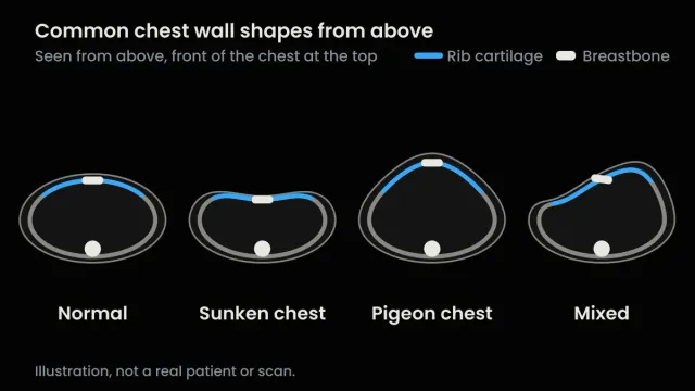
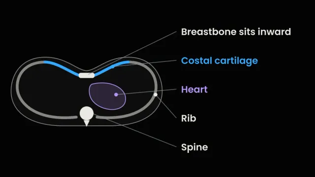
The difference comes down to position. In pectus excavatum the breastbone sits behind the line of the ribs on either side, usually in its lower half, so the chest dips where it would normally be level. Normal chests aren’t perfectly flat either: in one CT study, a third of children with no suspected chest wall problem had a small variant that radiologists called normal. A doctor’s exam decides whether yours is pectus. A scan measures how deep it is.
Got an X-ray report? Skip to pectus excavatum on an X-ray.
I was diagnosed with pectus excavatum at 15 and corrected it without surgery over about three years. The change was visible and measurable, but not complete. I’m not a doctor, so every medical point below comes from a named study or hospital page, my own views are labelled, and where I couldn’t find a study, I say so.
Normal chest vs pectus excavatum at a glance
| View | Normal chest | Pectus excavatum |
|---|---|---|
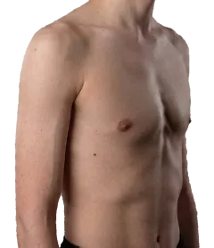
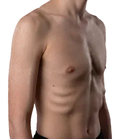
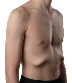
| From the front | Breastbone roughly level with the ribs beside it. Small tilts and bumps are common | A dip over the breastbone, usually low down and often right of centre. The lower ribs can stick out |
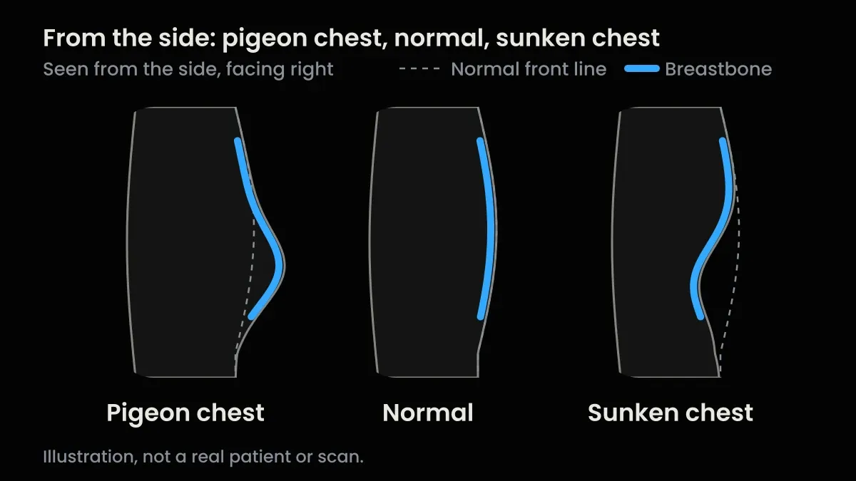
| From the side | The front of the chest runs fairly smoothly down from the collarbone | The breastbone sits behind the line of the ribs, and the lower ribs can jut forward below the dip |
| Front X-ray | Right edge of the heart visible, ribs curved like a C | Clues are often subtle: right heart edge blurred, heart shifted left, front ribs steep like a 7 |
| Side X-ray | Clear space behind the breastbone | The breastbone sits back toward the heart |
| CT or MRI | Haller index about 2.5 on average | A higher index (3.25 is a common line for a surgical opinion), sometimes with the heart pushed aside |
What a normal chest looks like
A normal chest isn’t a flat board. In a CT study of 200 US children with no suspected chest wall problem, 33% had at least one variant at the front of the chest. Most often it was a tilted breastbone or a rib or cartilage curving forward. The radiologists called these normal, and they were no more common at one age or in one sex than another. That’s children only. I couldn’t find a matching study in adults.
The scan number varies as well. The Haller index is a ratio taken from a CT or MRI slice (more on it below). In normal children it runs from about 1.9 to 2.7, according to a 2016 review of chest wall imaging in the British Journal of Radiology, and about 2.5 is the usual average quoted for a chest without pectus excavatum. In a Children’s Mercy Kansas City study, 220 people without pectus averaged 2.35, against 4.06 in 252 who had surgery for it, and the two groups’ numbers overlapped.
So there’s no clean line where normal stops and pectus starts. “Normal” is a range, and where a chest sits in it is a call for a doctor’s exam. If you’re a parent comparing your child’s chest, my guide to sunken chest in babies and children covers what changes with growth.
From the front
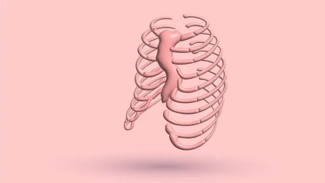
From the front, pectus excavatum is a dip over the breastbone (sternum). Cleveland Clinic describes the breastbone growing inward, with an indentation that involves four or five ribs on each side. The costal cartilage, the tough strips that join the ribs to the breastbone, goes in with it. It’s also called sunken chest or funnel chest. It’s usually there from birth, but people often notice it in their early teens. Cleveland Clinic puts it at about 1 to 8 people in 1,000, and estimates vary.
The shape varies. In 300 patients at one large US surgical clinic in Norfolk, Virginia, whose average Haller index was 4.9, so deeper chests than most:
- Cup-shaped, a localized deep dip: 67%
- Saucer-shaped, broad and shallower: 21%
- Trench-like, a long furrow: 10%
The deepest point was right of the midline in 80%, and by photo the dip was in the lower breastbone in 75%. Keep in mind these were people who came to a surgeon.
Don’t lean on the labels. When six chest wall surgeons in Buenos Aires sorted photos of 100 patients into these shapes, they fully agreed on only 30%. If specialists can’t agree from a photo, a mirror won’t settle the label either.
The lower ribs can stick out too, with the bottom edge of the ribcage poking forward below the dip. That has its own guide, flared ribs. The other signs are in pectus excavatum symptoms.
In women and girls, breasts can hide the dip and how deep it is, which my guide to pectus excavatum in women covers.
From the side
Side-on, the difference is clearest. In a normal chest, the breastbone sits roughly level with the front of the ribcage beside it. In pectus excavatum, the ribs on each side sit further forward than the breastbone, as the 2016 imaging review puts it. Lay a straight edge across the front of the ribcage and, in pectus, the breastbone sits behind it.
That leaves less room in front of the spine. The same review says this can push the heart to the left and rotate it, which I cover in pectus excavatum and heart problems.
The straight edge is my plain-words picture, not a measured rule. I couldn’t find a source that gives a normal breastbone angle or depth from the side.
The rest of the body can look different too, on average. Prof. Mustafa Yüksel, who reviews medical content on this site, co-authored a 2021 study of 90 teens with pectus excavatum, 90 with pectus carinatum and 90 healthy teens, aged 10 to 18, in Turkey. Both pectus groups weighed less and scored lower on posture and physical function than the healthy teens. How severe the chest was wasn’t linked to those scores. How the teens saw their chest’s appearance was.
A smaller 2024 study in Ankara (22 teens with each condition, 21 healthy) found more upper-back rounding in both pectus groups.
Those are group averages in teens, and they don’t describe any one chest. My view: posture changes how deep the dip looks, not how deep it is. Slump and it looks deeper. Stand tall and it looks shallower, and the breastbone hasn’t moved. My pectus posture guide has a two-minute check.
Pectus excavatum on an X-ray
A chest X-ray is a flat picture, so a dip that’s obvious side-on can be easy to miss. The front view is called a PA film (posteroanterior, because the beam passes from back to front), and the side view a lateral film. The classic clues:
- The right edge of the heart is blurred or hidden, sometimes with a hazy patch in the lung beside it.
- The heart sits further to the left, because the breastbone takes up some of the space in front of the spine.
- The front ribs look steep. They slant down “like a 7, rather than the normal C-shape”, in the words of the Radiology Masterclass teaching page by a UK consultant radiologist.
- The back ribs look horizontal.
- On the side film, the breastbone sits back toward the heart.
Before you check an X-ray against that list: a 2019 New Zealand radiology study says these signs “are often subtle and rarely all present in any individual.” A normal-looking front film doesn’t rule pectus out. The side view shows the breastbone directly.
Two clues trip people up. One is the hazy patch. In a case report of one teenager, the front film showed what looked like a denser area over the right lung, the kind of shadow that can be mistaken for pneumonia. The side film showed the breastbone pushed in, which explained it.
The other is the heart’s size. On a front film the heart can look enlarged, but part of that look comes from it being squeezed and shifted, as the Learning Radiology teaching notes explain. Looking big on an X-ray isn’t the same as being big.
Sometimes pectus is first spotted on an X-ray taken for something else. In a Dutch search of about 160,000 X-ray reports, mostly of older adults, it was noted in 1 to 2 per 1,000. That’s how often it was written down, not how common it is.
Women have an extra clue, from the same New Zealand study: sharper, more upright inner breast edges on the front film, called the medial breast margin sign. The women’s guide above explains it.
A Haller index can also be worked out from a front and side X-ray, at about 0.02 mSv (millisieverts, the unit for radiation dose). It’s a rough one, least reliable near 3.25 and in lopsided chests.
On a CT or MRI scan
A CT scan builds slices from X-rays. An MRI uses a magnet and radio waves, with no radiation. Both show the chest in cross-section, as if cut straight across, which is where the Haller index comes from.
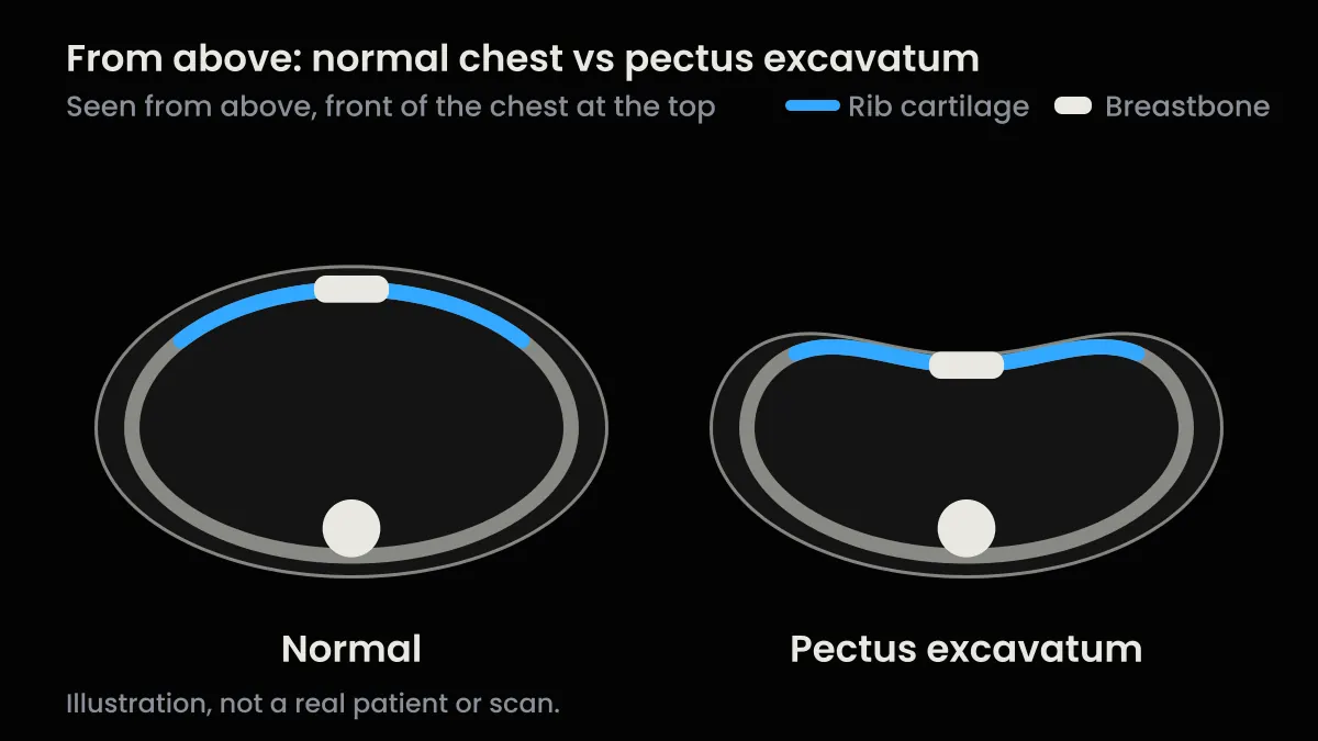
On the slice where the dip is deepest, the index divides the widest inside width of the chest by the shortest distance from the back of the breastbone to the front of the spine. About 2.5 is average for a chest without pectus excavatum, and 3.25 is a common line for a surgical opinion, but no single number decides surgery. What your number means, and why two scans can disagree, is in my Haller index guide.
This table draws mostly on a 2024 review of pectus imaging:
| Test | What it shows | Haller index? | Radiation | Worth knowing |
|---|---|---|---|---|
| Front and side X-ray | Clues on the front, breastbone position on the side | A rough one | About 0.02 mSv | Least reliable near 3.25, and not suited to very lopsided chests |
| CT | The full shape, heart squeezed or pushed aside, lopsidedness, a twisted breastbone | Yes, the reference | About 1 to 2 mSv, or about 0.35 or less with low-dose pectus scans | Radiation is the main concern, especially in children and teens. Often used to measure severity when surgery is being considered |
| MRI | The same shape detail, plus how the heart is working | Yes, similar to CT in a Beijing study of children | None | Less widely available, and costs more |
| 3D surface scan | The outside of the chest only | No | None | Promising but not standardized, and less accurate with breasts or more body fat |
No single test covers it all. A 2019 review by specialists in Buenos Aires and Spain gives each one a role: CT for how severe the shape is, cardiac MRI for where and how much the heart is squeezed, and a stress echo (a heart ultrasound during exercise) for whether that changes how the heart works.
The number also moves with how you’re scanned. It reads higher at the end of a breath out than a breath in, and standing matters too. In a small Japanese study using upright CT, 21 people with pectus excavatum (median age 22, 18 of them male) had a median index of 10.1 standing against 6.4 lying down. The dip was no deeper. The lower spine shifted forward when they stood. In 35 healthy volunteers it barely moved, 2.9 against 2.8. Scans are normally done lying down, so compare like with like.
And a ruler or caliper reading at home isn’t a Haller index. Skin-based ratios run on a lower scale, so use yours to track change over time and don’t compare it with a scan score.
Pectus excavatum vs pigeon chest
Pectus excavatum is the opposite of pigeon chest. In pigeon chest (pectus carinatum), the breastbone and the cartilage beside it push outward instead of in, so from the side the breastbone sits in front of the rib line rather than behind it. It’s the less common of the two, according to the 2016 imaging review.
It hides better on an X-ray, too: the same review says the front film is often unremarkable, and the side film shows the bump. On a CT, the Haller index works the other way round, with a lower number meaning a more severe pigeon chest.
You can have both at once. In the Norfolk clinic’s 300 patients, 1% had a mixed shape. In 430 children with pigeon chest in Southampton, 15 had a dip and a bump together.
For pigeon chest itself, see what pectus carinatum is and its side-by-side table. Mixed and rarer shapes are in chest wall deformities. And if what you’ve got is a bump rather than a dip, start with sternum sticking out.
Shapes that get mistaken for it
What separates them is where the change sits: the breastbone, the side of the ribs, the whole chest or the rib edge.
| Shape | Where it is | What it looks like | Usually linked to |
|---|---|---|---|
| Pectus excavatum | The breastbone, usually its lower half | A dip in the middle, with the breastbone behind the rib line | Usually present from birth, often noticed in the early teens |
| Harrison’s groove | Along the lower ribs, around the 6th, usually on both sides | A sideways dent running from the bottom of the breastbone out toward the armpit | Rickets, long-term breathing problems such as asthma or COPD, repeated chest infections, or no clear cause |
| Barrel chest | The whole chest | Full and rounded, as if holding a deep breath | Ageing and lung conditions such as COPD, asthma or emphysema, among other causes |
| A visible rib edge or rib flare | The bottom edge of the ribcage | The rib margin showing or sticking out, with the breastbone level | Can be normal in lean people. Flare can also come with pectus |
Harrison’s groove, as the medical reference StatPearls describes it, is a groove across the lower ribs, not a sunken breastbone, though it can turn up alongside pectus excavatum or pigeon chest.
For barrel chest, Cleveland Clinic describes a broad, rounded chest that looks like a barrel. My interpretation: it’s roughly the reverse of pectus excavatum. A barrel chest looks full and rounded, while pectus shortens the gap between breastbone and spine at the dip. Cleveland Clinic doesn’t compare the two.
When to see a doctor
See a doctor before you start any device, brace or exercise program if any of these apply to you:
- chest pain
- fainting or nearly fainting
- breathlessness at rest or with light effort
- palpitations or an irregular heartbeat
- a diagnosed heart or lung condition
- Marfan syndrome or Ehlers-Danlos syndrome, or a doctor has raised either
- previous chest surgery
- a Haller index above 3.25 together with symptoms
- symptoms that nobody has checked yet
None of these means you’ll need surgery. They mean someone should look at your heart and lungs before you go further. An index above 3.25 is a common referral threshold, not an emergency.
Even without any of these, a doctor’s exam is what tells pectus from a normal variant or something else. Cleveland Clinic says a simple physical exam can find it.
Who to see. Start with your family doctor. If they find something, or your chest is severe and you have symptoms, ask for a referral to a pediatric or thoracic (chest) surgeon who works with chest wall conditions.
What to do next
Pick the line that sounds like you.
- You want to know if it’s changing. Track it the same way each month with my guide to measuring pectus excavatum at home. Same photos, same ruler, not a guess in the mirror.
- You have a scan number. The Haller index guide linked in the scan section above explains what it means.
- You’re not sure how significant yours is. Take my free 1-minute quiz. It’s a quick severity check, not a diagnosis.
- You want the whole picture. Causes, symptoms and options are in what pectus excavatum is.
Comparing your chest with a normal one tells you what’s different. It doesn’t tell you how much that matters for you. That takes a doctor’s exam, and sometimes a scan.
Sources
- Donnelly LF, et al. Radiology. 1999;212(3):837-40. Read it
- Mak SM, et al. Br J Radiol. 2016;89(1061):20150595. Read it
- St Peter SD, et al. J Pediatr Surg. 2011;46(12):2270-3. Read it
- Pectus Excavatum. Cleveland Clinic. Updated 2023 Aug 11 [cited 2026 Oct 21]. Read it
- Kelly RE Jr, et al. Arch Bronconeumol. 2013;49(5):196-200. Read it
- Rodríguez-Ortiz D, et al. J Pediatr Surg. 2026;61(3):162872. Read it
- Alaca N, Yüksel M. Pediatr Surg Int. 2021;37(6):765-75. Read it Co-authored by Prof. Mustafa Yüksel.
- Mete O, et al. Pediatr Surg Int. 2024;40(1):178. Read it
- Chest X-ray, anatomical variants: pectus excavatum. Radiology Masterclass (Lloyd-Jones G). Reviewed 2019 Oct [cited 2026 Oct 21]. Read it
- Hapuarachchi KS, Cowan IA. J Med Imaging Radiat Oncol. 2019;63(6):737-44. Read it
- Robbins LP. Radiol Case Rep. 2011;6(1):460. Read it
- Pectus Excavatum Deformity. LearningRadiology. [cited 2026 Oct 21]. Read it
- Winkens RA, et al. ISRN Fam Med. 2013;2013:373059. Read it
- Coorens NA, et al. J Thorac Dis. 2024;16(1):696-707. Read it
- Sun J, et al. Pediatr Investig. 2019;3(2):102-9. Read it
- Rodríguez-Granillo GA, et al. Eur J Radiol. 2019;117:140-8. Read it
- Suzuki T, et al. Eur J Cardiothorac Surg. 2024;66(5):ezae408. Read it
- Bough GM, et al. Pediatr Surg Int. 2025;42(1):56. Read it
- Yoham AL, Sajjad H. Anatomy, Abdomen and Pelvis: Harrison Groove. StatPearls. Updated 2022 Oct 10 [cited 2026 Oct 21]. Read it
- Barrel Chest. Cleveland Clinic. Reviewed 2026 Mar 6 [cited 2026 Oct 21]. Read it
Reading about the condition is one thing. Knowing which one you have, how severe it is and what to do first takes about a minute.
- Your condition named, not guessed
- A severity read you can act on
- The exact next step for your case

This article is for educational purposes only and does not constitute medical advice. Always consult a qualified healthcare professional before starting any treatment plan. View full disclaimer →